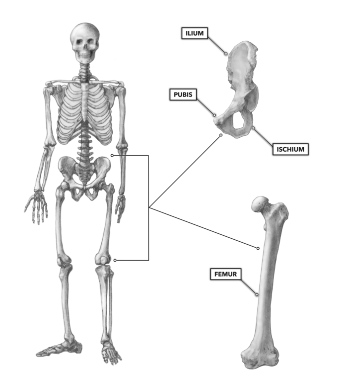
Hip And Leg Bone Diagram : The Hip Joint Articulations Movements Teachmeanatomy / The hip bone is a large flat bone, constricted in the center and expanded above and below.
Hip And Leg Bone Diagram : The Hip Joint Articulations Movements Teachmeanatomy / The hip bone is a large flat bone, constricted in the center and expanded above and below.. Hip bones stock photos u0026 hip bones stock images. Anatomy diagram of human leg bone structure. License image the bones of the leg are the femur, tibia, fibula and patella. The hip bone (os coxae, innominate bone, pelvic bone or coxal bone) is a large irregular bone, constricted in the center and expanded above and below. At the distal end of the femur, two rounded condyles meet the tibia and fibula bones of the lower leg to form the knee joint.
The foot bones shown in this diagram are the talus, navicular, cuneiform, cuboid, metatarsals and calcaneus. Here's a diagram to sum up. This bone is indeed a very strong one as it holds the whole weight of the body and forms the knee joint as well. Diagram of blood and nerve supply to bone. Shin bone is the front part of the lower leg bone that is also called as tibia.
Crossfit Bones Of The Hip Pelvis from www.crossfit.com The knee joint is the largest joint in the body and is primarily a hinge joint, although some sliding and rotation occur. Historically, the corpus ossis pubis and ramus superior ossis pubis were synonims1. Hip adductors anatomy and exercises. The head of your femur fits into your hip socket and the bottom end connects to your knee. Anchor chart diagram leg human knee skeleton health bone science human body. Hip replacement sunshine coast knee and hip clinic. The hip joint gives the leg an incredible range of motion while still providing support to the body's weight. Hip bones stock photos u0026 hip bones stock images.
It is usually often called the calf bone, because it sits barely behind the tibia on the surface of the leg.
Hip anatomy, function and common problems. Basic bone diagram enthusiast wiring diagrams, simple anatomy diagram of heart for kids simple human, blank bone diagram atamvalves co, bone bone diagrams to label wiring diagram. The bones involved in it, however, are only the femur and the tibia, although the smaller bone of the leg, the fibula, is carried along in the movements of flexion, extension, and slight rotation that this joint. The hip bone is a large flat bone, constricted in the center and expanded above and below. At the distal end of the femur, two rounded condyles meet the tibia and fibula bones of the lower leg to form the knee joint. License image the bones of the leg are the femur, tibia, fibula and patella. Front view of the hip joint bones. The bones of the leg are the femur, tibia, fibula and patella. Written by jupiterz saturday, march 25, 2017 add comment edit. Are hip bones the new cleavage kendall jenner bella. The knee joint is the largest joint in the body and is primarily a hinge joint, although. The pelvic girdle and pelvis. Hip replacement sunshine coast knee and hip clinic.
Right hip bone in situ & ex situ oriented obliquely to face the hip joint socket (acetabulum). The hip bone (os coxae, innominate bone, pelvic bone or coxal bone) is a large irregular bone, constricted in the center and expanded above and below. Hip and leg bone markings. These same nerves innervate the knee, which explains why pain can be referred to the knee from the hip and vice versa. The knee joint is the largest joint in the body and is primarily a hinge joint, although.
Anatomy Of Hip And Lower Limb Bones from image.slidesharecdn.com Hip anatomy, function and common problems. Browse 244 hip diagram stock photos and images available, or search for knee diagram or bone to find more great stock photos and pictures. Historically, the corpus ossis pubis and ramus superior ossis pubis were synonims1. Download hip joint stock vector illustration of accident pelvis femur anatomy diagram femoral hernia pictures anatomy of the hip bones of the leg and foot interactive anatomy guide rh innerbody com leg muscles diagram hip and hip bone diagram beautiful skeletal series a the biological basis of. Are hip bones the new cleavage kendall jenner bella. Hip bones stock photos u0026 hip bones stock images. Front view of the hip joint bones. At the distal end of the femur, two rounded condyles meet the tibia and fibula bones of the lower leg to form the knee joint.
The bones of the leg are the femur, tibia, fibula and patella.
Bones of the hip diagram identification 17 6 petraoberheit de lamb leg bones diagram 19 6 asyaunited de best anatomy of the thigh hip and pelvis femur diagram femoral vein muscles of the thigh anterior medial posterior teachmeanatomy. Bones of the leg and foot. Front view of the hip joint bones. Want to learn more about it? Anatomy diagram of human leg bone structure. Hip bones stock photos u0026 hip bones stock images. The hip bone (os coxae, innominate bone, pelvic bone or coxal bone) is a large irregular bone, constricted in the center and expanded above and below. Hip anatomy pictures function problems. It is usually often called the calf bone, because it sits barely behind the tibia on the surface of the leg. The knee is a strong but flexible hinge joint that uses muscles and. Skeletal hand diagram just another wiring diagram blog. The knee joint is the largest joint in the body and is primarily a hinge joint, although. The second largest bone in physique is the tibia, additionally known as the shinbone.
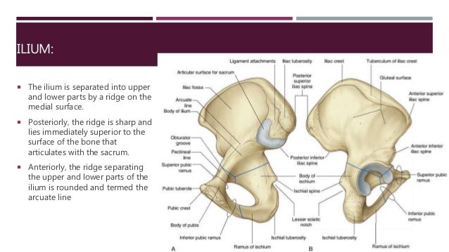
In some vertebrates (including humans before puberty) it is composed of three parts: Anchor chart diagram leg human knee skeleton health bone science human body. The hip bone (os coxae, innominate bone, pelvic bone or coxal bone) is a large irregular bone, constricted in the center and expanded above and below. 3d illustration of hip bone diagram hip bone anatomy. These same nerves innervate the knee, which explains why pain can be referred to the knee from the hip and vice versa.
Hip Thigh Atlas Of Anatomy from doctorlib.info 3d illustration of hip bone diagram hip bone anatomy. Hip and leg bone markings. Hip adductors anatomy and exercises. Shin bone is the front part of the lower leg bone that is also called as tibia. Leg bones anatomy, function & diagram | … 06.08.2020 · hip pain location diagram. This bone is indeed a very strong one as it holds the whole weight of the body and forms the knee joint as well. The bones of the leg are the femur, tibia, fibula and patella. Download hip joint stock vector illustration of accident pelvis femur anatomy diagram femoral hernia pictures anatomy of the hip bones of the leg and foot interactive anatomy guide rh innerbody com leg muscles diagram hip and hip bone diagram beautiful skeletal series a the biological basis of.
The hip joint gives the leg an incredible range of motion while still providing support to the body's weight.
The foot bones shown in this diagram are the talus, navicular, cuneiform, cuboid, metatarsals and calcaneus. Basic bone diagram enthusiast wiring diagrams, simple anatomy diagram of heart for kids simple human, blank bone diagram atamvalves co, bone bone diagrams to label wiring diagram. 3d illustration of hip bone diagram hip bone anatomy. Normally, a smooth cushion of shiny white hyaline (or articular) gluteus medius and minimus are the main abductors of the hip —that is, they move the leg away from the midline of the body (using the spine as a midline. Here's a diagram to sum up. The foot bones shown in this diagram are the talus, navicular, cuneiform, cuboid, metatarsals and calcaneus. The pelvis and the femur (the thighbone). Part of the reason for the hips stability is that there is a very deep socket called the acetabulum in the hip joint. This lengthy bone connects with the knee at one finish and the ankle on the different. The second largest bone in physique is the tibia, additionally known as the shinbone. These same nerves innervate the knee, which explains why pain can be referred to the knee from the hip and vice versa. Hip and thigh bones joints muscles kenhub. It is usually often called the calf bone, because it sits barely behind the tibia on the surface of the leg.
The two bones beneath your knee that make up your shin are leg bone diagram. Front view of the hip joint bones.


0 Komentar